| Product name | p21 Polyclonal Antibody |
| Immunogen | Synthesized peptide derived from human p21 around the non-phosphorylation site of T145 |
| Host | Rabbit |
| Reactivity | Human, Mouse, Rat |
| Applications | ELISA, IF, IHC-P, WB |
| Applications notes | Optimal working dilutions should be determined experimentally by the investigator. Suggested starting dilutions are as follows: WB (1:500-1:2000), IF (1:50-1:200), ELISA (1:20000). Not yet tested in other applications. |
| Clonality | Polyclonal |
| Preparation method | The antibody was affinity-purified from rabbit antiserum by affinity-chromatography using epitope-specific immunogen |
| Alternative | CDKN1A; CAP20; CDKN1; CIP1; MDA6; PIC1; SDI1; WAF1; Cyclin-dependent kinase inhibitor 1; CDK-interacting protein 1; Melanoma differentiation-associated protein 6; MDA-6; p21 |
| Formulation | Liquid solution |
| Concentration | 1 mg/ml |
| Molecular weight | 26 KD |
| Storage buffer | PBS containing 50% Glycerol, 0.5% BSA and 0.02% Sodium Azide. |
| Storage instructions | Stable for one year at -20°C from date of shipment. For maximum recovery of product, centrifuge the original vial after thawing and prior to removing the cap. Aliquot to avoid repeated freezing and thawing. |
| Shipping | Gel pack with blue ice. |
| Precautions | The product listed herein is for research use only and is not intended for use in human or clinical diagnosis. Suggested applications of our products are not recommendations to use our products in violation of any patent or as a license. We cannot be responsible for patent infringements or other violations that may occur with the use of this product. |
| Background | CDKN1A encodes a potent cyclin-dependent kinase inhibitor. Cyclin dependent kinase inhibitor 1A binds to and inhibits the activity of cyclin-cyclin-dependent kinase2 or -cyclin-dependent kinase4 complexes, and thus functions as a regulator of cell cycle progression at G1. The expression of CDKN1A is tightly controlled by the tumor suppressor protein p53, through which cyclin dependent kinase inhibitor 1A |
| Gene ID | 1026 |
| Alternative | CDKN1A; CAP20; CDKN1; CIP1; MDA6; PIC1; SDI1; WAF1; Cyclin-dependent kinase inhibitor 1; CDK-interacting protein 1; Melanoma differentiation-associated protein 6; MDA-6; p21 |
| Others | p21 Polyclonal Antibody detects endogenous levels of p21 protein. |
| Accession | P38936 |
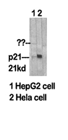
Fig.1. Western Blot analysis of HepG2 (1), Hela (2).
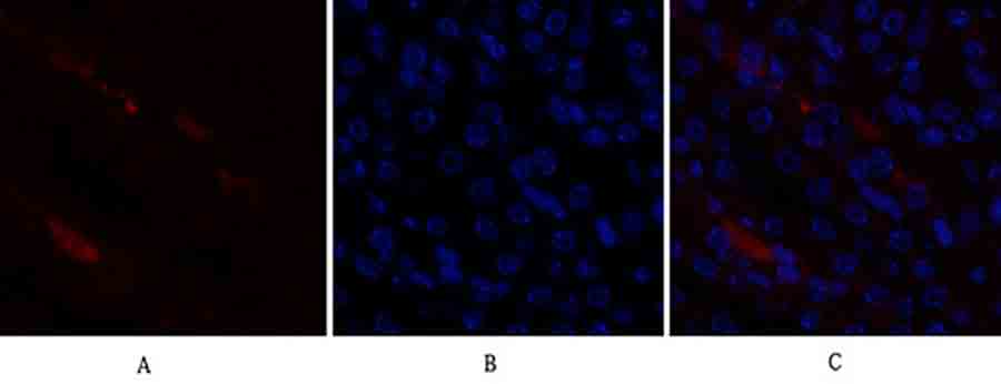
Fig.2. Immunofluorescence analysis of mouse kidney tissue. 1, p21 Polyclonal Antibody (red) was diluted at 1:200 (4°C, overnight). 2, Cy3 Labeled secondary antibody was diluted at 1:300 (room temperature, 50min). 3, Picture B: DAPI (blue) 10min. Picture A: Target. Picture B: DAPI. Picture C: merge of A+B.
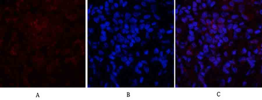
Fig.3. Immunofluorescence analysis of rat lung tissue. 1, p21 Polyclonal Antibody (red) was diluted at 1:200 (4°C, overnight). 2, Cy3 Labeled secondary antibody was diluted at 1:300 (room temperature, 50min). 3, Picture B: DAPI (blue) 10min. Picture A: Target. Picture B: DAPI. Picture C: merge of A+B.
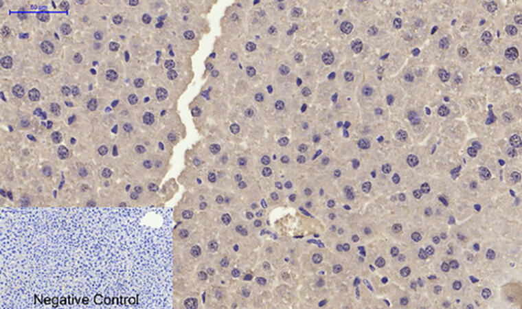
Fig.4. Immunohistochemical analysis of paraffin-embedded mouse liver tissue. 1, p21 Polyclonal Antibody was diluted at 1:200 (4°C, overnight). 2, Sodium citrate pH 6.0 was used for antibody retrieval (>98°C, 20min). 3, secondary antibody was diluted at 1:200 (room temperature, 30min). Negative control was used by secondary antibody only.
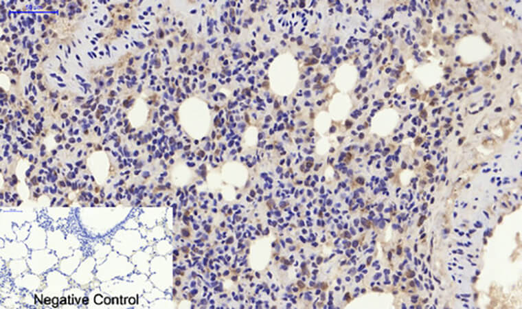
Fig.5. Immunohistochemical analysis of paraffin-embedded rat lung tissue. 1, p21 Polyclonal Antibody was diluted at 1:200 (4°C, overnight). 2, Sodium citrate pH 6.0 was used for antibody retrieval (>98°C, 20min). 3, secondary antibody was diluted at 1:200 (room temperature, 30min). Negative control was used by secondary antibody only.
You must be logged in to post a review.
1.The species of antibody reactivity should be the sample species that can be matched normally after Abbkine R&D experts have passed strict scientific verification. If your sample is not within the range of reactivity, in order to improve the efficiency and results of your experiment, it is not suggested to try other species. Otherwise, it may lead to sample mismatch and affect the effect of your experiment.
2.Please aliquot the antibody received as soon as possible and store it at -20℃, avoid repeated freezing and thawing, and use it within one year.
Welcome any form of communications, and better service will be provided here.
Tell: +1-404-854-0155
Email: service@abbkine.com
Support Email: support@abbkine.com
Address: 3052 Stroop Hill Road, Apt 203, Atlanta 30303, Georgia, United States of America


Reviews
There are no reviews yet.